Ultrasound Spleen with Dr. Alka Singhal | Medality / MRI Online Radiology Course
Watch on YouTubeVideo summary
The ultrasound evaluation of the spleen is a critical component of abdominal imaging, often requiring specific techniques such as a posterior approach with slight patient rotation to avoid obscuration by lung gas. Located in the left hypochondrium behind the stomach, this highly vascular organ receives its blood supply directly from the splenic artery without collateral flow, meaning any vessel blockage leads immediately to infarction. Normal splenic parenchyma appears homogeneous and hypoechoic with smooth outer margins, though measurement accuracy depends on visualizing both poles; while a length under 12 cm is generally standard, norms can vary based on age, body size, and population characteristics. The spleen serves as an important acoustic window for adjacent structures like the pancreatic tail, left kidney, and adrenal gland, and its drainage occurs via the splenic vein into the portal system.
Pathological conditions affecting the spleen range from congenital anomalies to infectious processes and malignancies. Congenital issues include accessory spleens, which are homogeneous lesions matching normal echotexture and can be confirmed via Doppler continuity with the splenic vessels, as well as wandering spleens that risk torsion due to lax ligaments. Infectious etiologies present with distinct features, such as pyogenic abscesses showing thick walls with posterior enhancement, fungal infections displaying a target appearance in immunocompromised patients, tuberculosis manifesting as multiple hyperechoic nodules, and parasitic cysts like hydatid cysts. Neoplasms vary from benign hemangiomas to malignant lymphoma or angiosarcoma, while metastases are rare but typically appear hyperechoic and avascular on Doppler imaging. Splenic infarction is characterized by a wedge-shaped hypoechoic area extending to the capsule, whereas advanced techniques like elastography and contrast-enhanced ultrasound help assess stiffness for portal hypertension and characterize focal lesions when CT contrast is contraindicated.
Traumatic injuries to the spleen are classified using the American Association of Surgery of Trauma (AAST) grading system, which distinguishes between lacerations defined as tears and ruptures involving a capsule burst. Grade 1 injuries involve subcapsular hematomas under 10% of the surface area or less than 1 cm in depth, while Grade 2 covers 10–50% surface area or hematomas up to 3 cm; Grades 3 through 5 represent severe damage with large intra-abdominal hematomas, vascular pedicle injuries, or multiple ruptures that often necessitate splenectomy. On ultrasound, these injuries appear as linear defects for lacerations and stellate patterns with irregular margins for ruptures, with contrast-enhanced ultrasound aiding in differentiating abscesses from malignancies by showing rim enhancement without central enhancement. Clinicians must also be aware of pitfalls such as mistaking the left liver lobe, interpositional masses, or adrenal tumors for pathology, and recognize that spleen size norms can extend up to 14 cm in individuals with large body habitus while smaller sizes may be normal in the elderly.
In conclusion, the comprehensive assessment of the spleen requires a nuanced understanding of its anatomy, vascular supply, and diverse pathological presentations to avoid misdiagnosis. While biopsy is rarely indicated due to the organ's high vascularity and associated risks, Doppler evaluation remains essential for tracing flow from the hilum through the portal vein and hepatic vessels, particularly in cases of chronic liver disease. The session emphasizes that accurate diagnosis relies on distinguishing between normal variants like accessory spleens and true pathology, utilizing both standard grayscale imaging and advanced modalities like elastography and contrast-enhanced ultrasound to guide management decisions ranging from conservative observation for low-grade trauma to surgical intervention for severe injuries or malignancies.
Read the full video transcript
Hello and welcome to noon conference
hosted by modality. Noon conference
connects the global radiology community
through free live educational webinars
that are accessible for all as an
opportunity to learn alongside top
radiologists from around the world.
Today we are honored to welcome Dr. Ala
Singal for a lecture entitled ultrasound
spleen. Dr. Dr. Singal is an associate
director of radiology at Madant Menic
City Hospital Delhi NCR India and has
over 28 years of experience in
radiology. She's authored several
publications and talks for leading
national and international conferences
and is the author of the Atlas of
Parathyroid ultrasound.
At the end of the lecture, please join
her in a Q&A session where she will
address questions you may have on
today's topic. Please remember to use
that Q&A feature to submit your
questions so we can get to as many as we
can before our time is up. With that we
are ready to begin today's lecture. Dr.
Singhol please take it from here.
>> Thank you. And so today's ultrasound
topic is ultrasound of the spleen. Now
spleen is a very it's a very simple
topic I think as we all would recall
during ultrasound what do we do with the
spleen evaluation? We simply see whether
it's there or not and what's the size
and if at all maybe is there any focal
pathology but trust me paying a little
more attention to it it's such a vital
organ to do the scan and if we do it
diligently we can actually evaluate the
eeology of the spleenic enlargement and
pathology
and let's understand how
so okay so it's often called the
forgotten an organ of the abdominal
scan. So we do a routine abdominal scan
but okay as we do liver we focus so much
of time attention for the liver
gallbladder pancreas kidneys and all the
other organs but spleen has not been
given that much attention however
recently so spleen elasttography and
contrast ultrasound and routine
evaluation is getting more and more
important and especially our imaging
technology has really improvised and the
way we look at this Explain the amount
of findings that we are able to observe.
In cases of diffuse pathological changes
and focal pathological changes both are
immense. So more is visible to the eye
more is being diagnosed on ultrasound.
Let's see. So of course like for any
other pathology any part of the body we
will be uh evaluating under continal
anomalies infections benign and neoplasm
and other non-neoplastic and
miscellaneous conditions right so
[sighs and gasps] let's see to begin
with of course anatomy defines
everything so we say spleen is the
largest organ of the lymphatic system of
course sizes vary and the healthy adult
spleen. So why we use the word is
healthy and adult and spleen because the
size varies with the population. So size
varies with the age, size varies with
the uh with the health of the patient.
So it's a very important window to the
direction in which the clinical uh
diagnosis is there lie. Where is it
located? As we all know it's located in
the left highondrium.
posterior to the spenic fracture in the
stomach making an interior approach
almost invariably unsuccessful due to an
overlying bowel gas because the stomach
is sitting right interior. So you do
need to rotate the patient a bit in the
cubitus position and scan from a little
posterior approach and that way we see
it nicely. A little
breath hold is very helpful but not too
much. That's we will understand why
because then the plural gas also
obscures it in the lung space. Now as we
all know spleen is a very vascular organ
and what's [clears throat] the blood
supply? It receives a blood supply from
the spleenic artery which is a branch of
the cedi axis and the intrappleenic
branches to north anastimos or
communicate with one another. There is
no intrplenic collateral flow. So what
that practically means that in fact
would be common in case of any blockage
to any particular vessel that area of
the spleen is rendered avascular and
under goes infact. So without collateral
flow the spleen is at an increased risk
for infection that must always be
remembered and especially we see a lot
of where I work a lot of CLT patients
with chronic liver disease and really
enlarged spleen and that's a common
cause of an intraabdominal emergency and
pain and presentation as well and the
spleenic vein the draining vein unites
with the superior mentric vein posterior
to the neck of the pancreas to form the
hippatic portal vein And evaluation of
the vascule is equally important to
exclude any thrombus and any other
aneurysm or any other pathology in the
arterial venus
vascule.
Right? So like we discussed spleen is
located in the left upper quadrant and
it's readily examined in several
positions. often you may need to really
work around and we will also know uh a
study in the flexibility or the mobility
study that we will do for wandering
spleen that we do need to rotate the
patient. We need to put the patient in
various positions to see if there is any
change in the position of the spleen
with the deicitus and and any shift in
the position if there is any one brain
splane and of course the transducer we
use the routine 3.5 MHz transducer
rarely for in children where very
superficial any abnormality any focal
pathology fungal infection when you
really want to evaluate the detail the
target appearance of
lesion then we can go up if we can go up
with a higher frequency transducer it's
definitely gives us more clarity and
more accuracy in the diagnosis
right so that's about the transducer so
approach like I said is the left lateral
and costal is always the best in supine
position and so the gas will bal is
usually interior to the spleen and
gentle respir respiration is frequently
more successful than the deeper
inspiration
because otherwise the lung bases come
downwards and obscure the spleen
altogether. So you would have noticed
beginners often find it challenging to
find spleen because of the over
inspiration at times. So the goal is to
direct that unobstructed ultrasound beam
between the lower intercostal spaces
towards the spleen along its midaxis. So
we can have the full cranio cordal
measurement of the spleen from upper to
the lower pole. So that's the
probe placement and that's the image
that you would be accordingly creating
and that's very uh supportive in the
accurate measurement. Right? [gasps] Now
what does this bean parent normally look
like? So as we all know it's like a
homogeneous
smooth diffused hypoquic eos right and
the shape it's very nice smooth rounded
outer margin and a nodular slightly
nodular indulating inner margin and at
times you may see accessory spleen as we
will shortly evaluate.
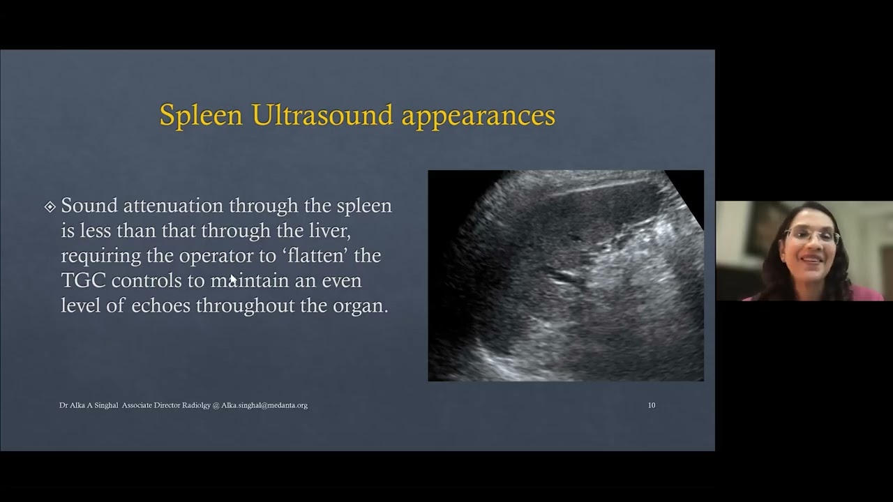
So also a difference between the spleen
and the liver is that you may you need
you need lesser TGC gain settings
because uh the t uh the there's not much
beam attenuation in the spleen as
compared to the liver and uh that's an
important tip to know and sometimes you
of course you may have droardia and
situs invertus. So the location of the
spleen instead of being on the left side
it may be on the other side. So of
course how do you identify the liver is
with the portal vein and the CBD inside
but if you look at the spleen there
wouldn't be any portal vein and CBD
there would be just hilar vessels which
are branching into the spleen parenchima
and each of these is an end artery there
are no inter
collateral or anastasmosis between them.
So to be able to differentiate between
the liver and spleen is also a important
task like not to misinterpret which
organ are you looking at right of
course. So so we will also whenever we
need to do the doctor studies we look at
the flow and diameters of the spleenic
vein and document them.
So just to orientation of the anatomy
when we describe the lesion. So we have
this is the outer surface as we can see
here at the laser pointer. Okay. So
that's the outer surface that's the
inner surface or the medial surface of
the hilum that's closely aparting the
pancreatic tail. So many times a window
to the pancreas the pancreatic tail can
also be achieved through the spleen as a
because that's a nice solid organ and
the pancreatic tail is sitting there.
Sometimes we can have pancreatitis where
we may not have much changes in the head
and the body but just in pathology in
the tail region or any uh intra intra
pancreatic accessory spleen or any other
pathology. So using spleenic uh uh
window to evaluate the pancreas is
another great ultrasound skill that you
can use. [snorts]
Right. So of course you'll see the
spleenic pain and you can see this the
upper pole that's the lower pole and
that's and that can also change
depending upon how you've labeled your
orientation in the ultrasound machine.
So obviously you will have to remember
that how you planned it all out but
that's the nature most people use. So
the vascule the Doppler we will always
study. So oh it's like I said the spleen
provides also an acoustic window to the
upper port of the left kidney. the left
adrenal and the tail of the pancreas. So
to evaluate these organs also this is a
very nice window to use.
Okay. So of course we can also
demonstrate the haler vessels. All
right. [gasps]
So the first and the foremost thing or
maybe for most people the only thing
that we do is screen is it there or not
and what does it measure and is there
any focal abnormality to let's put it
simple way and then first we see the
size and then we see the focal
abnormality. Let's first come to the
size. Of course, for the size, it
definitely comes you have actually
demonstrated the organ completely
because if you only get part of the
organ in your field of view and if you
measure that you'll obviously not be
documenting the full's complete size and
that would not be an accurate estimation
of the size of the spleen.
So going back to my previous image. So,
demonstration of a nice lower pole in
the image. Demonstration of a nice upper
pole in the image and then putting your
calipers on. So, I could actually uh
have a little wider sector width if
that's possible or allow that before I
can do my length measurement. Okay. So,
just to remember that. So, demonstration
of a nice upper and lower pole is very
important when we proceed with the
length measurement.
Okay. So of course spleen size and shape
are both variable like I discussed. So
what is the length of the spleen which
is the inferior the pole to the superior
pole and usually below 12 cm is
considered within normal limits but of
course we have to look at the age body
size of the patient. You can have a lean
thin young female and you can have a
large bodybuild patient. Of course, the
spreading size would be different,
right? Of course, the length is an
adequate indicator for most purposes and
provides a useful baseline for
monitoring changes in disease status.
So, routinely we only take one
measurement but 7 cm in transverse or 12
cm in the length is a upper limit of the
normal. Now the spleen as aging happens
it it goes down the size it increases in
the ch it is up in the childhood and
then it goes smaller in the elderly
the spleen enlarges downwards and
medially. So sometimes in chronic liver
disease especially so because the left
lobe of liver is also gone under
hypertrophy and the spleen is alo also
enlarged cranial cordally. So you have
to be and the ecotexture of the parent
uh may have a resemblance. So be careful
in observing which organ are you
measuring.
So of course spleen will grow downwards
into and it pushes the left kidney into
the pelvic often you will find that. So
that's like a spleen which is nicely
elongated. That's just the direction and
that's the placement of the caliber. A
nice demonstration of the dome of
diaphragm is very helpful and it is very
convincing that you have actually
included the whole of the spleen there.
Right? So that's when you see the hilum
then that is the point which is
confirming that you are if that's the
three-dimensional volume of the organ
that you are in the right plane to
measure that because many times you see
the spleen images which are not showing
the hila vessels and a measurement for
the longitudinal axis has been made that
would not be correct. So you must show
demonstrate the higher upper pole and
lower pole and then you do the length
measurement and that's the width
measurement. You can do the depth and
the width as well.
Right? So that's the length measurement.
That's again some certain examples. It's
a case of chronic liver disease where we
do see a lot of varices in the uh medial
part of the spleen. Another approach
which I don't adopt is what literature
says you could use a panoramic view or
extended field of view to measure the
length of the spleen especially in cases
of large spleen when you cannot
accommodate or include it all in one
frame. However, of course it there will
be a lot of ri shadowing and it's
technically a little challenging
especially because you need breath
holding and maneuvering of a transducer.
But yes, perhaps that's another option
to do it for those who are skilled with
their MSK work and really love to do
anoromic views. right now the spleen
size especially because in children uh
before 12 years of age so we we follow
normograms the ch uh the percentiles
like there are norm for the various ages
that if it falls within the 3 to 97% of
percentile that's the median splein
velocity so this is a paper that has
been published uh by uh by aims uh in
India. So that's the which is pertaining
to Indian population. So that we
routinely follow to evaluate for the
spleenic size as compared to the age the
median size and then we uh give the
dimension whether it's enlarged or not
enlarged
for a 5year-old, 6 year old, 7 year old
and of course it's also got the sizes
for the uh liver as well. Okay. Now, so
like we discussed what are the causes
for spleen omegaally. Now after we have
seen the spleen size of course when we
are evaluating any patient for an
ultrasound scan we are usually going to
be first looking at the clinical history
as why did the patient come to us? What
is the clinical presentation? So that
will obviously give us an idea as to
what are we looking at? Is there any
slee? Is there any s uh chronic liver
disease? Is there any renal disease? Is
there any infection? Is there any
gastroenterthritis?
So that will give you as a cue the
patient's running fever. Are we looking
at biogenic infection or what are the
causes? So the various conditions
causing diffuse enlargement of the
spleen including congestive hypoplastic
inflammatory infection and filerative
causes. So ultrasound will not be
definite but you can still give a
direction to the possible ethology
of the diagnosis right so broadly four
categories infection CD portal
habitation
or hematological disorders and
neoplastic conditions so let's look at
this in more detail
okay so broadly like I said the
categories are congestive causes as in
liver cerosis codal hypertension and
congestive cardiac failure. Infections
which could be acute infections such as
infectious monucleiosis.
chronic such as millary tuberculosis uh
inflammatory causes such as sarcoidosis
and SLE and then neoplastic lymphoma is
a very common cause leukemia and
metastasis and of course myop
proliferative disorders extra metal
hemopois as it's happening in
siri and storage diseases and many other
cases which will also lead to spleenic
enlargement
what do we see so if it's Spleenomeally
due to portal hypertension. Of course,
we will see associated viruses, dilated
spleenic vein, dilated spleen portal
junction and maybe varises all over in
the abdomen at the sitski sides in the
ligamentum ties leal ligament ligamentum
uh falsify ligament and vein and all the
other areas. So also what happens in
this the inferior margin is like a
rounded it's not like a sharp one.
descending below and medial to the left
kidney viruses are seen associated
changes of chronic liver disease would
be seen in the liver maybe pathologies
and so that you can see
[sighs]
right so we will look at more specific
causes as we come here now what about
the conal abnormalities of the speed
let's look at the big broad heading
because that we do come across in our
everyday scanning and these often cause
diagnostic dilemmas in unless proved
otherwise. Let's put it that way.
Simple. So what are these? Common ones
are the accessory spleen also known as
splitulus or a small spenic nodule.
Common sites are the spleenic hilum and
they could also be intraancriatic
like especially within the pancreatic
tail region. They could be in the other
sides the less common sides anywhere in
the mental. So intraancriotic accessory
spleen could be there and then we could
also have wandering spleen which is
basically hyper mortality of the spleen.
So we it's spleen
uh is seen in uh beyond the left
hypochondrium in the other areas in the
abdomen and of course that can also
enlarge that can undergo pathological
changes and that is also liable to
torsion and other pathologies as we
shall see soon right so let's understand
them each so accessory spleen of course
arises from a failure of the embriionic
spleen and arch to fused and then
extreme lovation that separates spenic
tissue is formed and this is very
common. So 10 to 30% of individuals
which is fairly common usually found
around the tail of the around the
spicilum or in the ligaments in the
pancreatic tail or sometimes in the
pancreatic parenime
ultrasound you will see well
circumscribed hypocoic homogeneous like
same equexture as this be the best part
is that's your reflection and the mirror
upon usually 1 cm or less or so maybe
but rarely seen more than 3 cm right so
that is the characteristic and of course
the common sites as I explained already
are the uh highlighter region then you
can have the tail of the pancreas and
less often you can see them in the
spinocolic ligament the greater momentum
misentry anywhere in the pelvis as well
right so uh the percentages commonly of
course is 75% is spinic high and the
rest the lesser lesser percentages at
the other sides.
How does it look like? Like we see. So
in here we have a this is the normal
spleen and that's the well definfined
rounded hypercoid well circumscribed
area. Echogenicity is quite matching
that of the spleenic paraga that is the
confirmatory that is the schematic
diagram to show how an intraaran creatic
accessory spleen would look like. So a
welldefined hypocoic nodular lesion
which is in the spleen. So it's not now
the our commonest differential of course
here would be a pancreatic mass a
pancreatic primary pathology
and definitely uh we would be doing a
contrast CCD abdomen to prove it or a
contrast ultrasound to prove it but on a
gray scale a lesion that looks as
similar ecoenist as of spenic parenta
can be uh the spenic spelankulus is in
the differential diagnosis.
another case of a pancreatic uh
intraarent pancreatic
uh splankus that we can see here.
So the why do we need to observe and
document them because they also enlarge
under the same circumstances which cause
pedome and they were also hypertrophy in
postpinctomy patients. So observing
making a note of them and documenting
health in differential diagnosis and
giving explanation to find and seen on
imaging.
So we can use our Doppler and the
circulation of the splankus derives from
the main splenic artery and drains into
the main splenic vein. So when you trace
the blood supply that can help you in
differentiation. And what is the
importance? Importance is of course not
to mistate them from lymph nodes, not to
mistate them from an adrenal lesion and
not to mistake them from a mass in the
tail of pancreas. So conversely, so if
it's a mass in the tail of pancreas and
you mistake as a splankus, of course
that's not going to be a good news. The
left adrenal mass and you mistake it as
spraining, that's also not good. And
there's a lymph node enlargement in the
abdomen that may be required in
sampling. You cannot label that as plas.
So these three differential diagnosis
are very important and of course each
has their own characteristic
identification. Lymph nodes would have
their high vascularity and hypocoic
rounded appearance that you can
identify. Adrenal nodule will have its
either it's a lipo
lipoid or hypocoic or some hetrogenous
parenma or typically located at the
upper pole. the shape and other
pathology will give you a cue. Mass in
the pal of the pancreas again you
observe where the vascule and the
feeding is coming to it. A splenulus
will derive its vessel vasculature from
the spleenic artery. So if you evaluate
and patiently
scan you may be able to demonstrate the
connectivity and give a differential
diagnosis.
Next coming to wandering spleen. Now the
name sounds very weird. Spleen is going
on wandering or it's ectopic in location
or where it is. So basically what this
refers to when the spleen migrates from
its normally fixed posterolateral
location in the left upper abdomen to
other areas in the abdomen. So condition
can be both conidal and acquired. And
why it happens? because there's there's
a lack of normal anchoring by the
gastrosplenic and the gastroenal
ligaments.
So ultrasound is a valuable modality to
diagnose this and to evaluate its
complications including if there is any
torsion or impaction in that case right.
So of course in torsion and infaction we
will look at infaction which will come
up in later and torsion you'll see
always a twisted pedicle and loss of
vascule or supply in the organ and
you'll see demonstrate a twisted pedicle
and a world cool sign right of a lot or
something and in fact you'll see a veg
shaped area hypervascular zone right uh
watershed uh area. So in particular most
important is like a realtime ultrasound
helps test the spleenic hypermobility
and the vascular cortical abnormalities
right now. How do we test for spenic
hypermobility? Let's see. So basically
you have the patient in leftus. The
spleen normally just stays there on the
upper side. Right? But if the spleen
gravitates to a dependent area on the
right side of the abdomen or to the
pelvis or to any other area that means
there is not much anchorage with the
gastrosplenic and the lead recommends
right. So unlike in the static stem so
what happens is you position the patient
in the right decipitus observe the
spleen physically migrating to the
dependent positions within the abdomen.
So this by this we can track the
spleen's gravitational shift and its
migratory nature and we can confirm if
it's absent from its normal quadrant and
it's freely moving. Right? So two things
you will need to establish that it's
actually left its quadrant and it is
moving. Okay? Right? So this is a case
where we see the spleen which is uh uh
which is uh not a nice pointed rounded
shape. It's like a flattened shape and
it's enlarged and citrogenous
and it's descending down and it's uh it
presented with acute abdomen and there's
torsion and eskeeia of the wand screen.
So
uh we have to evaluate the vascularity
right treat uh diagnosis is confirmatory
is by CCT abdomen and treatment is by
spleenopaxi which is uh reposition the
spleen and to prevent the portion and
pre preserve its function.
So that was about the cenital anomalies
right. Moving on to the next a section
of abnormalities that we see very
commonly for which we are actually
sought to evaluate as to what it is
especially fungal infections we see very
commonly not very commonly but we see in
transplant recipients or imunompromised
patients. Biogenic absesses in spleen
are less frequently seen as compared to
liver. we see more uh amic abapsis and
uh other absesses but you can see them
in the screen as well. Tubercular
millary tuberculosis and tubercular
absis again in endemic areas can be
seen. Of course parasitic infection and
hydrated cysts are very common in
endemic areas and of course in Asia they
are common right. So pyogenic absis like
anywhere in the body you can see
solitary and multiple variable thick
sized or anoic hypericid cyst without
any flow. Same for fungal infection
you'll see small target shaped like
nodules microapsises less than 1 cm
usually tubercular are small meary
nodules and parasitic hydrated cyst
would have variable all stages depending
upon what stage of the hydrated cyst you
are looking at clear or with folding
membranes undulating membranes floating
sand or a calcified hydrated cyst right
let's understand in details so what do
we have here we have Bacterial
infections we have spenic abscesses for
bacterial infections we what are the
common causes
infectious spread is usually through the
bloodstream 75% cases or through trauma
or through spenic infaction.
So a rendered avascular area is of
course susceptible to infection right.
So like we discussed uh it can be single
or multiple. If we have poorly defined
onto hypergoic or any cystic masses of
course like an absess like a cyst the
characteristic as we've discussed in
ultrasound basis there's a posterior
acoustic enhancement and of course we at
times post intervention or otherwise you
can see some air bubbles and shadowing
as well. So basically a hetrogenous
ragged margins hypercoic lesion with
some peripheral vascularity that may be
seen definitely aspiration and relieving
the pressure definitely supports in the
healing. You can have absesses with some
septi and loilulation as well and
aspiration release the fever
symptomatology and escurative
fungal infections usually are seen in
immunompromised people. Again you see
these hypoquic nodular areas. However,
if you observe them closely they will
each show a central echogenic dot as
well. So that's what's we call a target
appearance. like we see an echogenic
area. So this is about the layering
pattern. So fungal infections typically
occur in imuninompromised people and
with neutropenia and commonly caused by
candida aspergillosis and cryptocus.
Fungal infection appear as microapsis
usually less than a cm
and almost always less than 2 cm.
Candida can show bull eye or wheel
within a wheel pattern consisting of
layers of necrosis, inflammation and
fibrosis. Right? See here this we there
are some absesses in the spleen uh in
the liver as well along with multiple
absesses in the spleen.
So this is characteristic appearance is
the target appearance. You can use a
higher frequency transducer to
demonstrate this classic picture. So
that way you know that you're not
looking at me nodules or any other
sarcoidosis nodules or any other
pathology.
Coming to next tuberculosis.
So microacterial spleenic involvement
can develop in disseminated milary
tuberculosis. Of course what do we see
on ultrasound is multiple hypercodules
absesses can form and of course there is
spleenic enlargement associated with
this. There would be liver enlargement.
There would be multiple abdominal lymph
nodes and you can haveitis
you can have gerital tuberculosis and
findings in various other parts of the
body. So basically what you will see is
hypercoic nodules in the sple and which
are demonstrated both ultrasound and CT
and associated other findings in this
abdomen.
Next you can also have sarcoidosis which
is basically a diffused bleeding
enlargement and again mentric lymph
nodes FNA and other imaging is
diagnostic.
All right. So you can have illdefined
nodules which often just show up. Right.
Now coming to the more important area of
hydrated cyst. Basically parasitic
infections. Commonly we are concerned
about hydrated cyst because hydrated
sphenic hydrated liver hydrated is
another one of the indications for liver
transplant and we do keep on seeing them
very frequently.
So
uh which shows that it's still
communicable and it's uh it's still uh
endemic in certain parts of the world
and uh what we see on ultrasound of
course it depends on the stage of the
cyst and the associated complications.
What you see is anoic fluid fil cyst and
if you see any debris floating any any
indulation in the internal membrane
floating membranes any sand sometimes
you can see very classic like you know
picture appearances
really really this is like a hydrated
cyst you can see the cyst and you can
see the floating indulating membranes
with it again the floating indulating
membranes that you can see you sometimes
you can see classes the daughter's
chicises and the sand and all the other
appearances within. So all these are
classic appearances and u of course
these patients will require treatment
uh to prevent uh the disease from
spreading and localizing it.
So other cysts that you can see which
has just simple cyst to epidermoid spin
cyst or any other simple cyst calcific
or any other cyst you just have to
document them and uh they may not be
belonging to any specific infection or
any other category but yes documentation
is required
and um rarely they may undergo secondary
hemorrhage or infection and then you can
have internal debris and thicker walls
right
and Other parasitic infection could be
par
gonmiais where again you can find
hetrogenous solid h solid cystic
hypooric areas. Again this diagnosis
would be non-specific on ultrasound and
it's predominantly a hystopathological
diagnosis.
Right? So along with that hydrocyst and
other paracetic diseases would often be
seen spread in the abdomen. Right? So
you can see nodular pathol
in other areas of the abdomen as well.
Okay. Now coming to the neoplasms.
So the benign neoplasms the commonly
that we see him in tuma which we see and
don't see kind of the simple common less
than cm typical hypercoic nodules that
we see which are incidental. Of course
we see atypical heimomas where we are
the lesion is uh appearing hypercoic and
shows maybe some vascularity some
features and of course the CT is
required to confirm and exclude other
pathologies and then we know it's a hema
that's been labeled and then it's
constantly coming to us for for a
followup.
We can see hematomas where we see
homogeneous iso to hypercoic lesions
which are hypervascular. We can see
lympangoma. The classic appearance of
lymphoma is the echogenic lines that we
see in any part of the body multilocular
cyst. And if there are any soft tissue
nodules they may show some vascularity
and let's see
so the commonly the heimas is simple
that we see commonly soft small soft
tissue hypercoic areas similar to what
we see in the liver and larger lesions
may undergo
may be seen like this. Again this is the
lesion but of course triple phase CT the
flow characteristics confirm the heava
diagnosis
hematas
so rare it's basically an appearance of
a sleen within a spleen what it says is
it's malformed spiny tissue caused by
post-traumatic scar nodular hyperplasia
or hypoplastic nodules typically appears
as a single well-defined homogeneous
isoquic or hyperquic nodule mass or a
mass on ultrasound. So the key finding
is there is hypervascularity in it is
which is reflected as a hypervascular
pattern on ultrasound and it is
confirmed on cross. Look at the intense
enhancement and intense hypervascularity
of the lesion. Right? So that's
confirmatory. Now coming to the next the
lymphopolerative disorders. Lymphoma of
course is the most common malignant
disease and approximately 3% of spleenic
malignancies are lymphas.
So what is lympantioma? So benign slow
growing continal neoplasm seen in
childhood. Basically you see a
multilocular cystic lesion located in
the subcapsular area of the spleen may
contain intraocular intraocular ecoenic
debris hyperquid septations and thin
neural calcifications. Right? So that's
the kind of classic appearance that you
can see like a honeycomb or a
sponge-like anyway spaces and of course
size and followup is required. No
treatment as such is given. So you can
see them as multiloculated cystic mass
and these can be big huge as well.
Right. Coming to the next entity which
is SA antiscent sclerosing angtoid
nodular transformation. So basically
it's a nonoplastic vascular lesion which
shows multiple angic metal nodules
interspersed with hyper strand. So ba uh
you see vascularity
in the nodule and of course you can only
suggest a diagnosis confirmed by CT
right. So a hypoic nodule the classic
vascularity pattern is demonstrated
either on a contrast ultrasound or on a
contrast CCT abdomen.
Coming to the next the malignant masses
the lymphoma metastasis and angio
saroma.
So spenic lymphoma as we say is the one
of the commonest malignancy diffuse
infiltration you can just have spenic
enlargement or you can have focal
nodular pathology. Often you can see
along with enlargement illdefined
hyperacquic nodules throughout the
parenma.
You can also have focal
nodular masses in the spleenic parenma.
So,
so you can have meary nodules. So,
differentiation between milary
tuberculosis
again is clinical or hystopathological.
Coming to the next clinic metastasis.
Metastasis is uncommon, rare. However,
lymphoma is common. And other
malignancies that metastasize to spleen
are melanoma, cancers of breast, ovary,
colon,
lung and prostate. So most metastasis
occur as solitary or multiple nodules
while approximately
50% are hypercoid. This varies according
to the primary tumor and hypercoidic
especially from melanoma and colon
cancer mixed eco cystic and target
appearances are also seen most of the
metastases
are avascular on color after ultrasound
there isn't any vascularity that we are
able to demonstrate on ultrasound right
so and associated findings for
malignancy you will see metastasis
elsewhere in the body by the time you
have metastasis So that kind of
supports you in confirming the diagnosis
or giving a hint to as to what you're
looking at. So majority of the colon
metastasis and others are hypercoidic.
Coming to next angio saroma which is a
primary uh non-hemopoic malignant
neoplasm of the spleen develops from the
endometrial lining of the blood vessels
on the spleen. So it's seen as well
defined hetrogenous solitary or multiple
nodules of masses with hemorrhage or
necrotic changes and spenomeally
interusative cases. So you can
demonstrated an increased blood flow in
the solid component but not always
right. So coming to the next the cystic
lesions the cystic lesions they what
develops in the spleen they could be
just simple congenital cyst there could
be some pseudo cyst and in fact that
developed as an in fact to begin with
but later can become necro and cystic so
that could also look like cyst and then
of course you can see gamma antibodies
and piosis
very commonly seen spinic in fact. Now
what does it look like? The classic
appearance is if the speed that's the
outer surface you'll have the veg the
base of the triangle is at over there
and the triangular the aets is usually
at the hilum right so that kind of an
orientation if you find a W shapeology
that really looks like a triangular with
a base
towards the capsule outer capsule of the
spleen and vascular connectivity with
the spelic kila that is quite quite
suggestive of a in part right so
underlying hemolytic anemia as inle and
other pathologies cods and other things
are predisposing liations ultrasound
changes like whenever the in fact has
happened it takes some time before the
paranka alters for you to be able to
appreciate the difference the in the uh
and the line the watershed line right
so you do see a wedge-shaped hyperquake
area and a lack of flow and it can give
a mass-like appearance, irregular
mahogens or a not multilotular mortal
appearance in that scenario diagnosis
can be a dilemma right and chronic
stages uh or long-term it shrinks
ecoenity increases and there can be a
fibrosis issues right so this is a case
of spina that I just recently saw so you
see that's the
spleen and you see this This is the
capsular lesion which is got the base
over there and it's like uh reaching up
to the hilum. So it's a classic in the
central part of the spleen we have a
wedshaped hypergoic hetrogenous area
which is really characteristic of a
spleenic impact. So again you can see
there is a
capsular contour that is dropping at the
sight of the impact. So you can
classically demonstrate that yes that is
of [clears throat] course that's the
spleen measurement that's been enlarged
and that's the size of the inf 3 into 4
in 3 cm that's in the other orientation
that we demonstrate. We attempt to
demonstrate the blood vascularity. You
see some flow outside but not within the
area that you're talking about. So that
kind of confirms the diagnosis, right?
You can see.
Okay. All right.
So that was another case. That's a
different case. Another case where we
have in fact the whole of the lower pool
area here.
Gamma candy bodies. Commonly we see tiny
ecoenic bright dots cedotic not as
bright as it's seen in the image that's
a very classic picture. So of course you
can see in various uh situations of
congestive hematomey
cle cell disease hemolytic anemas
lemadoma hemocromattosis blood
transfusion and various other
situations.
So tiny dot-like calcifications that you
can spot on
spinic piliosis which is rare pathology
and is characterized by cinosodal like
dilotation formation of multiple cyst
like cavities within the parenma
so rarely seen in the liver common in
the uh common in the liver less often
seen in the spleen but that's what it
looks like basically you have poorly
defined hypergoic nodules again it we
can only give differential diagnosis
some calcified spin cy that you can see
they these could be hydrated we don't
know but we just document them the way
we see in fact we've already discussed
what are the causes that you can see we
can see there's a band sign like the
echogenic strand this hypergoic lines
that we see in the impact tissue that is
classic of in park so if you can
identify that then you don't need to
really think on the lines of an S you
you know it that you're dealing with an
impart see this kind of a white ecoenic
line appearance
that's that's the
appearance of the uh coral snake
right so you can identify the spenic in
part coming to the last two topics
little bit of spleen elastigraphy and
contrast ultrasound so of course
elastigraphy of the spleen is coming up
more and more and uh of course uh more
meta analysis is needed before we can
actually understand that how much it can
predict but yes it has a place in
chronic liver disease in predicting the
complications and um it is being uh done
we doing a research work on the liver
and spleen elasttoraphy together as a
non-invasive tool to assess the
complications of
uh CLD uh including viruses and other
parameters and correlating with lab
parameters.
So because the spleen is highly
vascular, stiffness closely reflects
changes in pressure throughout the
abdomen. So what is the rule? Primarily
of course diag spleen stiffness
measurements are highly accurate and
often outperform liver stiffness
indirect measurements and they support
in predicting isopial viruses. there
support in monitoring the liver pipe
process. So
how do we do it? There are we can do it
shear or we can do it as an RP tool as
an absolute velocity measurement. So and
we can give the measurement in kpa the
kilopascal or in meters/ second. And of
course beans uh stiffness values are
higher
um in cerosis and total hypertension
compared to normal population. So what
it says is that of course it's an
operator dependent
modality and of course reproducibility
is a question at the moment. However, it
is a promising tool and uh it may it may
add value to the esophagoscopy
and screening modality for detecting the
size and the presence of esophasial
viruses.
Coming to contrast ultrasound, we do a
lot of work on liver liver contrast
ultrasound. However, screen contrast of
course uh is usually honestly uh sought
when there is an impaired renal function
and we do not want to give a uh CT
contrast. So we can use ultrasound
contrast as another uh diagnostic tool
to evaluate any focal nodular pathology
of which we want to assert in the
pattern. The pattern is the same as
evaluating a focal lesion for the liver
and the enhancement pattern for any
infectious biogenic absis would be
peripheral enhancement for any
cysts simple cyst they do not enhance
for any uh melant nodules they hyper uh
they show rapid enhancement and rapid
wash out. So with that uh background
understanding we try and use the CUS for
lesion characterization.
Benign lesions typically show
enhancements similar uh to the normal
spenic tissue hypoenhancement
uh is seen with them. Trauma is a very
important role for this. So you see um
uh you can identify lacerations and
subcapsular hematomas and active
bleeding right at the bedside in
unstable patients when you can't do a
CT. So there is a great role of CUS in
trauma infactions there is a great role
in predicting and of course in benign
lesion excluding benign lesions and
leaving them the way they are. So heas
and hematimas we can see uh this just a
summary of the key points of course
which we've already covered and what I
wanted to cover now is the spleen trauma
injuries are common uh from sports and
various other factors you instantly the
patient is first sent for a vast scan so
you see fresh blood and absence of free
fluid however does not rule out any
spenic injury and we have to evaluate
the spenic
texture any laceration or hypergoic
regions presenting hematoma may be
present. So laceration is basically a
tear and rupture is rupture of the
capsule and hematoma is just a hematoma
for a peric capsular hematoma. So of
course there are five grades by the
American classification. So grade one is
a small subcapsular hematoma, grade two,
grade three and grade four. We'll come
to that. So various causes are uh most
common is of course trauma but a
predisposing condition of enlarged
spleen due to various causes is really
increases the vulnerability uh to
rupture. So basically what do we see on
ultrasound you can see there is some
altered eco texture it's not smooth. So
why is that happening? That is because
of an edematous tissue and there could
be subcapsular hematoma lacerations with
branching stellate appearance like
liberally shattered or a torn organ
hemoperonium fluid with eos that you see
now the American association of surgery
of trauma has created this devices
system which uh we will just learn and
laceration is a tear and rupture is a
bursting of the organs's outer capsules.
So that's what's a laceration. It's just
a sheer tear in it. And rupture is like
a rupture of the capsule at a focal
point. Right? So what is grade one?
Subcapsular hematoma less than 10%
surface area or less ration less than a
cm. Grade two is 10 to 50 cm% of surface
area hematoma or hematoma less than one
or less ration less than 1 to three.
Then grade three these are the ones
which are three four and five often
requires celctomy where is there is an
intraarent primal hematoma more than 5
cm or more than 50% surface area has
hematoma and of course grade four and
five have got injury to the vascular
picle or there's sheer multiple ruptures
of so here what it looks on ultrasound
you see a lesseration here you see a
lesseration and you see that that's what
you see on color At rupture you see
there's a sh there's a te like a
stellate like a star kind of an
appearance the capsules irregular
margins that you can see we can do
contrast a word so you can indicate it
in various studies you can see lesions
that don't enhance or that's the normal
enhancement that you see arterial and in
the venus phase there's a arterial phase
you'll see the early arterial vascular
enhancement and then in the venus the
space there's a homogeneous diffuse
enhancement of the organ. So that's just
how piogenic absis is presenting on a
contrast ultrasound. So it's enhancing
on the rim and there's no central
enhancement. So we are wanting to
exclude if it's a malignancy or a
metastasis but that's not what it is. So
it's just a uh pyogenic absis. So to
conclude remember the pitfalls of
course. So like I talked about the left
lo of liver like I talked about the
introspic masses like I talked about the
spinuli like I talked about the normal
tail of the pancreas and a left adrenal
mass. So remember these do not mistake
them for vice versa. And really I thank
you. I really really thank you all for
your presence for all your attention for
all the learning for creating this great
space. Thank you MRI online. I'm really
really grateful to all of you. Thank
you. Thank you so much for that lecture
Dr. Singhal. That was great.
We don't have any questions in the Q&A
box yet, but I can wait for one minute
to see if something comes in.
>> Sometimes it takes a second.
>> Hope you all like it and get some value
out of the session. The out would be
great. And yeah, do put in your
comments. So I do see some
appreciations. Thank you for the great
talk and insightful session. Thank you
so much. That is wonderful. And yeah,
okay you thank you for your presence and
listening.
Uh there's one thing how large can be
piosis?
I haven't personally seen but the
literature uh
would say probably I would say 3 to 5 cm
that would be my my guess. I haven't
seen large leations as such.
Thank you
and yeah
thank you for your question. So next is
when do you cause pleenomeally? I call
like especially if it's a child uh
depending on what age it is I follow the
I follow the pediatric uh paper that I
talked about uh sent out by as all India
institute of medical sciences that's
that's what most people here use for our
Indian population
uh that's the median percentiles that we
use so we call it more than 12 cm
because because otherwise in a normal
healthy adult, we don't normally see uh
12 plus or 13. There's usually some
chronic inflammation or something to
match up to. 14 is not more than 14. I
would call it more than 12.
[clears throat]
So yeah that would be my answer to the
question that you put in the chat
by Annabel and yeah
so please share us your experience on
biopsy or efficacy of spicy lesions.
So rarely is it sought as a diagnostic
dilemma to solve? Why would you biopsy a
knee lesion when you can't treat when
you really need a sample to treat the
pathology or you can't get a diagnosis.
So example if you have fungal infections
multiple fungal absesses so you may
require a samples to if you want to
tailor match the antibiotic
and if you have a like a say hydrated
you wouldn't put a needle in if you are
thinking of any
uh malignancies or uh there would be a
systematic systematic uh presentation
that will hint you to the diagnos nosis
much before you really need to put a
needle in uh in spleen for a sampling
and of course do remember spleen is a
highly vascular organ. So FNA of a
spleen is less frequently done and it's
less frequently required per se. I would
say put put through we can always sample
if it's required.
Uh says normaly in adults is 12 to 14.
Yeah because 12 to 14 it's written
because sometimes if you if I have a
very large built patient so I may give
it a consideration like I said. So for a
normal a median or a lower median build
I usually take it as 12 but if I have a
really large body habitus I would let go
14 up to 14 also as normal. So again I
think that's where the differences come
to because spleen size is really age and
body h and you know the distribution of
the population and the BMI index. It's a
lot factors to be accountable for.
Uh so question is can it be smaller than
normal? Can a normal spleen not be
visible due to gaps?
uh question of course uh spleen can be
smaller in elderly population spleens
that I you would see in lean thin small
build 70 year old 80 year old you might
just find barely a 5 cm or a 4 cm kind
of a spleen. So that is very normal
finding and can a normal spleen not be
visible due to gas? Yes, of course, it
can be obscured by balgases. That's why
I really paid attention on the scanning
technique for that. So, do not over
inspirate and hold uh just a light
breathe to just push the diaphragm a
little bit low so you can see it but not
push the lung too low so that it itself
secures the spleenic window. So that's
the intention. So if you remember that
it will make it simple.
Question for purposes of annual checkup
med life age person if length is 12 cm
you sign out as normal size. If length
is 12 up to 12 I wouldn't uh I wouldn't
quote it as abnormal.
So normal uh if it's more than 12 even
if it's 12.5 I'll call it as borderline
spleen.
And of course if it's more than 13 I
would label it as spin vigory. This is
how I use in my everyday practice.
Thank you. So next is thank you Dr.
Single. Is it possible to detect spenic
artery dilotation calcification
abnormality before the hilosone? Yes of
course the vessels are there. So that
that is actually a part of chronic liver
disease doppler. So then we do evaluate
the vessels very well. So we have to
take the vessels right from the
spicilum.
Uh the doppler evaluation for CLT starts
to evaluate the artery and the vein from
spenic highum to the SPJ then the porta
and you evaluate the hippatic artery and
the portal vein and all that cell. So
that's the hippatilary dropler. So it
includes the spleen at the far end and
the vascule and then the SPJ and you go
into the liver and the branching and the
hypatic veins and portal vein and the
hypatic artery. So yes of course you can
evaluate that. There are some more
questions in the chat. So question is
yeah I think a lot of gas that we've
covered and we'll check up. Yeah I think
we have covered the questions both in
the chat and in the Q&A. Yes.
Thank you.
>> Thank you so much. Yes, you got
everything. I appreciate you staying on
for a couple minutes and getting those.
Thank you so much for your lecture, Dr.
Single. We always love having you here.
And yes, thank you for everyone for
participating in this new conferences
conference and asking such great
questions. You can access a recording of
today's conference and all our previous
noon conferences by creating a free
account. We will also email out a link
to the replay later today. Be sure to
join us next week on Thursday, July 16th
at 12 p.m. Eastern where Dr. Omar Neimar
will deliver a lecture entitled benign
and malignant breast calcifications. You
can register for that at medality.com
and follow us on social media for
updates on future new conferences.
Thanks again for learning with us and
have a great day.