Synchrotron powder diffraction for the characterization of pharmaceuticals
Watch on YouTubeVideo summary
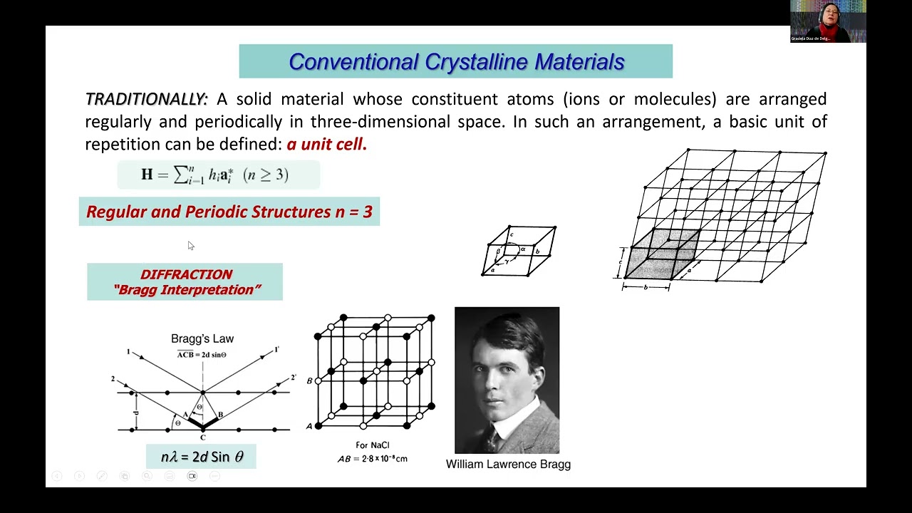
Dr. Gracilla Diaz from Universidad Los Andes in Venezuela presented an overview of synchrotron powder diffraction as a critical tool for characterizing pharmaceuticals, highlighting its essential role throughout the drug development lifecycle. She outlined four historical milestones in X-ray diffraction, beginning with the discovery of X-rays and progressing to the pivotal work on single crystals and the subsequent development of powder diffraction methods by scientists like Albert Hull. The presentation explained the fundamental principles behind these techniques, including Bragg's Law and the concept of reciprocal space, which allow researchers to determine unit cell parameters and atomic arrangements from diffraction patterns. Dr. Diaz emphasized that while conventional laboratory instruments are useful, synchrotron sources offer superior resolution and intensity, enabling the analysis of complex materials where preferred orientation or low crystallinity might otherwise obscure structural details.
The core applications discussed focused on identifying crystalline phases in poly-crystalline aggregates, quantifying their ratios, and analyzing microstructural features such as crystallite size and defects. Dr. Diaz detailed the standard workflow, which involves sample preparation using capillaries to minimize preferred orientation errors, followed by data collection at high-resolution beamlines like ID22 at ESRF in France. She explained how diffraction patterns serve as unique fingerprints for materials, allowing for qualitative identification against databases like PDF-5+ and Cambridge Structural Database (CSD). When unknown phases are detected, advanced algorithms such as DICVOL are used for indexing, followed by structural determination and refinement using programs like SHELX or JANA, often incorporating Density Functional Theory (DFT) calculations to validate the resulting atomic models.
To illustrate the practical advantages of synchrotron radiation, Dr. Diaz presented case studies involving active pharmaceutical ingredients like rasagiline mesylate, used for Parkinson's disease treatment, and various valproic acid derivatives. In these instances, laboratory data failed to yield correct structures due to significant preferred orientation effects that distorted peak intensities. However, by utilizing synchrotron data, the team successfully modeled these orientation effects accurately, leading to the determination of the correct crystal structures with high confidence. The presentation also covered temperature-dependent studies on calcium and strontium valproate derivatives, revealing complex phase transitions between monoclinic and triclinic structures that were only observable with the enhanced precision provided by synchrotron sources.
In conclusion, Dr. Diaz stressed that X-ray powder diffraction is indispensable for ensuring drug quality control, detecting counterfeit formulations, and resolving patent disputes regarding polymorphism and salt forms. She addressed audience questions regarding software accessibility, noting that tools like GSAS-II, DASH, and EXPO are available free of charge for structural analysis, though commercial packages often provide more automated interfaces. The session highlighted the collaborative nature of this research, involving institutions across Venezuela, Colombia, France, and Germany, and underscored how synchrotron facilities bridge the gap between theoretical models and experimental reality in pharmaceutical science.
Read the full video transcript
speaker Dr. Gracilla Diaz deal from
Univers Los Angeles in Venezuela and
she's going to make the presentation
singleton powder defraction for the
characterization of pharmaceuticals.
So we're very glad to have you with us.
Dr. Garcia, the floor is yours.
>> Thank you very much Karolina. I'm going
to share my screen. Start sharing my
screen.
I hope you can see it.
Presentation is preparing now. We can
see it. But uh in presentation mode
okay
>> I think it's opening
then now we can see it in presentation
mode. Thank you very much. You can get
ahead.
>> Okay. But I don't see it. I don't know
why.
We just uh
>> sometimes with F8.
>> Okay.
>> It can be shared as well.
>> Okay. Yeah, I went to to the other
screen.
>> Okay. I think I now I can see it also.
>> Fantastic. Go ahead, please.
>> Yeah. Thank you very much, Karolina.
It's a pleasure for me uh to participate
uh this time um as an instructor in this
very prestigious school. I will try to
to
summarize a little bit of uh what we
need to uh to keep in mind for uh polit
of a lamp. um
award uh as a fast team some years ago.
Uh but first I would like to acknowledge
uh my collaborators, my closest
collaborators,
professor Miguel Delgado and Professor
Analio Dugarte from the University from
University Los Angeles here in
Venezuela. Dr. Robert Toro and Professor
Jos Antonio now from Univers Industrial
Deantere in Bucharam Colombia, Andy Fish
and Kathine De Joy from ESRF in France
and Jakov and Distri from Avanguard uh
in Germany. I think we have been working
as a very good team in recent years and
uh we are all very glad to be to be able
to to share some of the work that we
have been doing recently.
Okay. And uh also because uh it's the
first time that uh I make a presentation
of this type, I will just uh like to
show you very briefly where I am
located. Um I am uh in Merida uh far
from the capital actually closer to the
border with Colombia.
Merida is one of the oldest states and
the university uh is has three campuses
and the main campus is in Merida. It has
two other campuses in in the in the
other Andian states of Venezuela. The
city is in a race valley at about uh
1,600 meters above sea level and we have
um usually we have a little bit snow in
the mountains and uh this is a uh view
of the of the city. erh the city the
university started as a seminary in uh
1785 and later became a university in
1810 soon after the declaration of
independence and then it became
universes.
Our faculty of sciences was founded in
1967.
Uh this is a view of the building and uh
some of the mountains and uh our lab
starts uh here uh from here. Uh so we
have a I think we have a good laboratory
space. Our laboratory was founded in
1969 and since then we have been um
trying to carry out the best possible
work with the with the resources uh
available um uh to us.
Uh the university is not the city is not
only a university city but it's also
very touristic uh place. It's very uh
well visited by uh Venezuelans and uh
people from abroad. And one of our major
attractions is the highest and the
second longest cable car system in the
world. And this is some of the
h typical views that uh we enjoy around
here. mountains with a little bit of
snow and the very emblematic uh
Spelleladia plant that it uh grows above
3,000 mters uh in this uh region. Okay.
So I I would like to start by pointing
out uh four milestones in X-ray
defraction. Of course we can uh think
about uh different uh many different
contributions uh but uh we think that uh
there are basically
four milestones that that need to be um
that that need to be uh remembered. Uh
first of all of course the discovery of
X-rays in 1895 by William Conrad Rangan
who obtained the Nobel Prize in physics
uh for his discovery.
The second one, the discovery of X-ray
defraction by crystals in about 1912.
Um, this was an experiment carried out
by Walter Fredick and Paul Nippen. Uh,
but the the experiment was suggested uh
by Maximon Lae. Maximum Lowi received
the Nobel Prize in chem in physics but
uh he shared with Fredick and Nippen uh
because of uh they they did uh a very
nice uh experimental work. Certainly a
major contribution was the defraction by
single crystals uh uh made by the by
William Henry and William Lawren father
and son that also made them um
recipients of the Nobel Prize in physics
uh this time in 1915
and uh
pretty much at the same time around 1916
in Europe
Peter Dubai and Paul Sher
and in the US Albert Hall developed the
the methods for
powder defraction uh using X-rays.
This is a very very important
contribution because uh of the um the
nature or or the the application that
powder diffraction has had over the
years uh in uh the characterization of
materials and we are going to emphasize
in this uh talk uh some of the most
important aspects uh of this uh of this
technique.
Let's first remember that what uh we
consider to be crystalline materials. H
for a crystalline material we need a
periodic a regular arrangement of atoms
and molecules h in some kind of
repetition pattern in three dimensions.
Uh we call conventional crystalline
materials to those uh compounds that
have a regular and periodic arrangement
of those uh atoms and molecules. When we
have an irregular and a periodic um
arrangement of those motives, we have
the so-cal amorphos materials. There is
a very important group of materials that
are regular but aperiodic
among which we find the quasi crystals
and Dan Shengman received the Nobel
Prize in chemistry in 2011 uh because of
the description of this uh of these
materials. uh but we are not going to uh
uh talk about this uh type of compound
which are very interesting but we are
not uh going to deal with them in in
this talk. So basically what uh we have
or what we uh would like to have is uh
some kind of a structural motive which
could be atoms or molecules or a complex
an organometallic complex that is
repeated according to a particular
pattern that is called the crystal lis
and uh to obtain
a regular arrangement of those motifs
atoms or molecules in three dimensions
that uh that arrangement can be uh
described uh in terms of a small
parallel pipet which is called the unit
cell. Erh that uh when we reproduce that
uh small unit with a a a unit that uh is
characterized by the smallest volume
possible and the highest symmetry
possible. when we repeat that uh that
unit in three dimensions we uh reproduce
the complete um crystal structure.
So um in traditionally in we consider
crystal uh conventional crystalline
materials those materials that are
that have a con constituent atoms or
ions or molecules in a regular and
periodical three-dimensional arrangement
in the in the space. uh in and in that
uh arrangement we uh define what is
called a unit cell that is uh described
in terms of uh three vectors and the
angles between them and when we repeat
this basic unit we uh reproduce the
whole crystal structure. Erh
one way to understand the interaction of
X-rays and actually the radiation with
uh an arrangement of this uh kind is uh
through the interpretation that Bra that
William Lawrence Brah gave to the
defraction phenomena. Of course there is
a very formal and very um
more um elaborate uh erh ways to to
present this but uh I think for the
purposes of uh this talk and for a a
general understanding of the of the
subject uh the brack interpretation is
very very convenient. So what uh Brack
proposed, William Lawrence Bra proposed
was that uh if we have a a set of um
X-rays of X-ray beams that um are in
interaction with the
planes, crystalline planes that we can
define in the in that unit cell.
Uh they will be defracted or in a
similar way as a reflection of light by
a mirror. they can be they will be
reflected with the same um angle of
incidence. H then we we can register
that um reflected uh beam. Uh but at the
same time there are many other erh rays
that are um interacting with the same uh
with with that particular set of planes.
So um some of them will be in phase
partially uh in phase or completely out
of phase.
The for those h emerging beams that have
a path difference which is an integer
integer number of the integer multi
multiple of the wavelength then we have
met the defraction condition. those uh
rays will be in phase and we will
produce a a reflection uh coming from
that particular set of planes that are
separated by a distance d
we can see that h
also in a simplified way here we have a
set of planes of crystalline planes that
uh are interacting with an x-ray beam
that it's make to
hit
those particular planes with an ang at
an angle of theta and when the the
defracted beam emerges also at that
theta angle we have a reflection. Those
planes are identified with their millar
indices HKL which are uh described in
terms of the inverse of the intercept of
that particular plane with different uh
important crystalallographic direction
in the in the in the structure. Here we
have um an animation of what happens and
uh we can see that when the the
uh to obtain uh a reflection the H rays
have to be in phase so that Bra's law is
uh follow is obeyed
depending on the way we have the sample
we may have a single crystals or we have
an aggregate of poly crystalline
material. In the case of single crystal
defraction, um we have just one
fragment, one single crystal that uh we
use um that uh we locate in the X-ray
beam and uh a series of reflections from
the different crystalline planes in that
structure um are produced and registered
in
nowadays in uh 2D detectors. So we can
have hundreds to thousands of
reflections produced by the interaction
of that X-ray beam. Actually can be also
neutrons and electrons with a crystal
that is been used uh in the experiment.
In politraction we have an aggregate of
a large number of very very small single
crystals. And uh also we make uh we we
use um an X-ray beam that uh interacts
with the specimen now composed of many
uh very small uh single crystals and um
what uh what is produced is a
one-dimensional or we what is registered
at the end is a one-dimensional
um
erh result
pattern resulting pattern which uh
contains a few maxima and some of those
maxima
um may be superimposed with reflections
from different uh sets of plane. So
that's one of the main characteristics
of the of the powder diffraction path.
One way to understand the the defraction
phenomena is the interpretation that was
proposed by Peter Paul Peter Ael based
on the in on the um construction of the
reciprocal lis and the eel sphere and
the idea that Bra's law is um obeyed is
um
holds under certain geometric
conditions. For example, if we have uh
the the the
cartoon that we had in the previous
slides where we have a set of uh
crystalline planes and I apologize
because uh this uh these names are are
in Spanish but we have a set of
crystalline planes identified by their
indices HKL and we have an incident
X-ray beam. We have a reflected beam at
um the same theta angle.
If we consider that the center of that
sample
is uh in a circle that is defined by the
inverse of the wavelength that is used
in the experiment
then it can be shown mathematically
here. I'm not going to um describe it in
detail. It can be shown that uh every
time that that emerging beam touches the
circle which actually by rotation it can
be made a sphere touches a sphere
constructed in this way called the evil
sphere then a brack law is obeyed and we
obtain a a defracted beam that we can
register with uh one uh available
detector. So the idea is to to have a
latis constructed h based on the inverse
of the spacing between the different set
of planes the so-called reciprocal lis
and that lis in some way is rotated to
make the different um
planes
um that describe that lattice touch the
sphere of reflection and at At that
point Bra's law is uh obey and we can
see a reflection. So with the positions
of those maxima the which are the the
theta hkl we can calculate the spacing
between the planes that are producing
that particular reflection and we can
from there we can calculate the unit
cell parameters of our uh crystalline
material. This is a another
uh uh small movie representing
uh how the different uh defracted uh
X-ray beams are uh obtained when we
bring the reciprocal lice to touch the
surface of the H sphere constructed in
the way that Eval
proposed. So we have a characteristic
pattern like this one that we register.
This is actually a single crystal
pattern but something similar happens uh
in powder diffraction. Basically what we
need is to have an X-ray source uh some
gometer somehow
of reflection. somehow a a detector uh
and
all these uh components um controlled by
a system that can uh
bring different crystalline planes to uh
follow the defraction condition that is
uh governed by by Bra. So that's the
basic uh thing with the defractometry.
H in that sense the direction of the
beam is important because it will give
us the size and shape of the unit cell
and the intensity of the beam is
important because it will it is related
to the nature
uh of the atoms and molecules that are
producing that reflection and the
special position um of of the different
of the different atoms in those planes.
So at the end that would be the way to
obtain the crystalline of structure of
that material. We have different types
of resources the conventional old X-ray
tubes uh more recent um sources as micro
focus and different types of uh
arrangements. Uh we have uh synretron
sources. This is a view of serious
synretron in Brazil and a view of ESRF
in Grenobyl. And we have also some other
emerging technologies like FELS that uh
promise uh and have already demonstrated
that are very useful in very um
particular and important cases for the
way to bring all the different uh planes
uh to follow the defraction condition.
we uh need somehow a a way to bring
those planes and uh that's why uh there
different uh geometries of gonometers
have been devised. This is um these are
two gonometers from uh different
commercial
uh vendors and but they use the brain
geometry which is the most common one in
in a laboratory setting for the
synretron. Uh this is a view of uh the
Hodgej at uh IBAM um ID22 beam line at
uh Grenoval.
And uh here we have uh a view of the of
the defractoter, the detector,
uh some uh device to uh for
uh low temperature analysis, a robot to
put the samples etc. Here, here we have
a more recent view of this gonometer
with a with a newer detector and we have
uh here um an amplified view of the
sample in a capillary mounted for
synretron data collection.
As for the detectors, we have uh point
detectors, linear detectors that are
useful, but uh of course they give a
view a limited portion of the of the
reciprocal space, but uh are um
nevertheless uh useful. And we also have
area detectors where uh we can collect
in a more rapid way several reflections
at the same time.
H basically the information contained in
a in a powder erh defraction pattern erh
we can uh visualize it here.
Erh
first of all we have the position of the
maxima which have to be recorded in the
best possible way and are related to the
unit cell the size and the shape of the
unit cell that describe that uh the
particular arrangement the particular
crystalline arrangement of one or more
crystalline components uh in the samp
We also have the relative intensities of
those reflections that is related to the
type and the position of the atoms in
the crystalline arrangement. We have the
shape of the maxima that is related to
the microructure. For example, the the
different um crystalline domains
changes in the shape due to tensions uh
defects uh in the structure etc. And we
have the background that includes a lot
of uh information which is nevertheless
uh important because it could indicate
the the presence of um amorphous faces,
the presence of diffuse scattering
and other type of um
low um crystallinity components.
For example,
uh one of the erh major problems uh that
uh we can um that we can encounter when
uh we use polifraction
uh particularly the brament geometry is
the it's the presence of several sources
of errors and for structural
determination from p diffraction the
most important one is the preferred
orientation
and uh this is something that uh that
needs to be uh taken well care of. Uh
for example, in the preparation of the
sample, a correct preparation of the
sample and uh but and there is uh also
ways to um to try to assess that uh
through some different algorithms. Uh
just to show a little bit of what um
what it looks like
if we have uh no uh preferential
orientation in a sample which is uh the
the
ideal situation. Uh all planes are
detected equally and we have a true
registry of the a a good registry of the
intensities of the of each reflection.
However, if we have uh a preferred
orientation, if the crystallites tend to
orient in a particular way as it's shown
here,
then uh we could have um
an effect on the intensities of uh
certain reflections that come from
different uh planes. So that's something
that is very important and is very
critical in braano geometry. Here
uh just to to summarize some of the most
important applications of X-ray powder
diffraction. We have uh the
identification of different crystalline
faces in a sample. Quantification of
crystalline faces and the structural
analysis of uh different materials which
has become more and more um erh easier
to to carry out. uh because of uh
different advances in instrumentation
and in the programs uh that deal with
this type of uh of information. So um
the most common application of
politraction is the identification of
crystalline components of components in
a poly crystalline aggregate.
And uh in this case uh we have uh a very
uh a nice review article uh in public
defraction. Here is the the link um on
the contribution of Albert Hall
in the US.
uh the most common application I mean
it's it's very important to identify
correctly the components of a poly
crystalline aggregates and this is also
a representation of that uh of that
particular case for example we have for
silicon dioxide several faces one is um
amorphos and we have other faces with
different crystal structures and the the
power of the
polraction methods is that uh it allows
us to identify each crystalline phase
because each crystalline phase has a
characteristic pattern and also in a
mixture of crystalline faces each phase
produces a a particular pattern that is
independent on the other fa phases. So
it allows not only qual um
identification qualitative
identification but also quantification
of the presence of those different
components.
The identification process uh can be
summarized here. Each powder diffraction
pattern is characteristic of a material.
So it is a we can say that is a
fingerprint and uh uh we can identify
several basic steps. We have a search um
of the defraction pattern uh usually in
a database a superposition or inhouse or
commercial databases. A superposition of
that particular experimental pattern
with a reference pattern that is found
in a database. The identification
deciding if the superposition of those
patterns is appropriate and if they
correspond to a a phase that is uh
already um published and repeat the
process for any other phases that are uh
present and at the end if possible
quantify the presence of uh each of the
crystalline phases. For that we have uh
very uh good databases uh in particular
for powder diffraction. The PDF 5 plus
database is the the database to use for
the identification of materials. The
Cambridge structural database contains
mostly single crystal structural
determinations but um
powder diffraction patterns can be
simulated from single crystal data and
they can also be used in conjunction
with the PDF5 uh plus database to
identify materials. Uh there is also the
COOD um database which contains a more
limited number of phases and uh in some
cases uh the quality of the of the
patterns is is not uh uniform across uh
across the database but uh it may be
useful for some applications and for
some uh researchers.
So um the powder diffraction file is the
the I would say this the the standard to
use to identify a powder um poly
materials in a poly crystalline sample
and uh it started you know as a simple
book as a simple registry by hand at the
beginning and became um after 1978 as
the ICD uh poly defraction file.
As I mentioned before, the COOD er
database is a open database that has uh
very good patterns for some materials.
Um
and uh here is a summary. I'm not going
to go into those details, but this is a
summary of the power of the powder
diffraction file for the
characterization of materials and the
Cambridge structural database uh which
um even though it is a basically a
single crystal powder defraction single
crystal uh file or database can be used
to obtain and here I have um highlighted
the programs and the algorithms that are
used in the
that can be used in powder defraction.
So the circular analysis uh using powder
defraction data may uh involve for
example
the identification of uh known faces. H
if we have erh after registering the
powder diffraction pattern we can
identify the crystalline phases using
the powder diffraction file or or the
Cambridge uh structural uh database or
the ICSD also the inorganic chemistry
database and uh if we have if we detect
an isosstructural phase use that phase
use the so-called SIF file in a in a
structural refinement
of that phase using several programs
some of them are public domain and some
of them are have a a cost but uh
nevertheless they they are all
important. Erh if we have unknown faces
then we have to go through an indexing
procedure using uh we we like uh dick
pole um
the the program uh dick bowl to
determine the unit cell parameters then
we need to do um having a step-by-step
profile of the pattern do a pattern
fitting and other decomposition
procedures um we can use the several
methodologies is there that are um
properly um reference to. We need to um
assign somehow study the systematic
presence of reflections to assign the
space group and uh we can carry out a
structural determination process using
uh different programs that use different
algorithms for example Dash Expo Janna
and other other programs that are
available. the structural refinement
usually uh by the rhythm method and uh
using the programs that we mentioned
before
the structure refinement uh using a
rhyth
summarized here I'm not going to go into
the the detail here uh because the
lecture will be um recorded uh but uh
this is uh basically what we have to do
to obtain the unit cell the nature and
the position of the atoms that will
describe the crystal structure. Erh
identify if there is more than one
component. Identify the quantitative
ratio of all those components and if um
necessary if appropriate um study the
microructure
which is determine the crystallite size
and any microructural
defects that might have uh that might
the the sample may have or the specimen
may have. And here we have uh tried to
combine or to present what would be the
the workflow
uh for um for a powder sample uh to go
from sample preparation, a sample
mounting either in a in a flat uh sample
holder or in capillaries as it is used
in mostly in the synretron and high
resolution. synretron uh radiation
collect the defraction pattern. I try to
identify the faces using the available
databases. If there are no entries on
those databases, then we need to go
through a an indexing profile feeding.
uh try to use some uh previously known
uh information on the on the molecule or
use a geometry optimization programs to
try to define um a structural model to
start with. Go through a structural
determination program
process uh using uh different methods
that are uh shown here.
structural refinement and uh hopefully
determine the crystal structure from
posraction data visualization. There are
several programs uh some are free, some
are uh with the license uh for
visualization,
validation and the FD calculation is
also very important and different uh
software for analysis of the material
like platon the mole that allow for
example the assessment of whether the
distances and angles that have been
obtained in that structural
determination procedure are
valid or not they if they make sense or
not and of course different types of uh
studies for of intermolecular
interactions.
These are some key references uh some uh
very important books uh that uh we need
to keep in mind but the key reference is
the volume age of the international
tables for a x-ray for
crystalallography.
uh and here is a summary and outline of
uh all the different parts. Uh so this
is a very important and very key
reference uh for powder diffraction. Uh
there is also a very nice um review in
nature review methods primers by a very
uh experienced group of um powder
defractionist.
Okay. So I'm just going to use a few
minutes to um to talk about some of the
work that we carried out at uh ESRF at
beamline ID22
with Andy Fish and Katherine De Joy
between in October to December of 22 and
in 2023.
uh we receive a grant and Alio and I uh
received a grant to through the LAMP uh
project to carry out a study and we took
to ESRF some of the problems that we had
encountered in our uh daily work that we
could not uh uh solve uh satisfactory
with laboratory uh powder defraction
data
Just to remind what's the interest of
pod defraction in the pharmaceutical
industry
at different points within in the erh
research and development or in the
production stages of a particular drug
it is important to characterize it by
different methods but x-ray defraction
is very very important in the
characterization of pharmaceutical
compounds. uh for example from the R&D
viewpoint uh at the laboratory stages we
need to characterize properly the
different active pharmaceutical
ingredients determine if they if they
present polymorphism if they are uh
obtained as salts or hydrates or any
other kind of sulfate um and any other
uh structural um feature that may be
important for the character
for the characterization of that
compound at the production level. It's
also important for quality control of
the of the drugs. H the analysis of the
raw materials, the exipients, fillers,
any kind of dice or any other type of uh
additional material that will go into
the formulation. We have to make sure
that it's the correct uh a API and that
exipients will not um
erh transform the material into
something that will be potentially
harmful uh to the to the patient that is
taking them. And uh the analysis of the
commercial product, the final commercial
product is also very important. It's and
it's also carried out with uh among
other techniques with X-ray powder
defraction or with in general powder
defraction erh to detect any kind of um
changes in the API
monitor the stability of the active
pharmaceutical ingredient any kind of
physical or chemical change in the
product aggregation change in color for
example which is not uh very uh not very
uh good to have occur in a medication
that we are uh using. Okay. Erh, it's
also important to determine the illegal
or counterfeit formulations uh something
that is uh unfortunately common in in
certain uh parts of the world. Uh and
also erh problems with the patent
infringement and things that have to be
that have to do with the litigation
processes uh because one company claims
um to own the patent to that particular
uh drug or formulation. And so it it
could be also a complicated thing. that
uh pol defraction actually is very
important to uh try to solve those kind
of problems.
So one of the erh one of the
er compounds uh one of the APIs that we
studied that we took actually to the to
the synretron to to try to h solve the
problem that that we have in the
structural determination was railene
meilate which is a a compound uh used in
the treatment of Parkinson disease. is
very very common and for in Latin
America in particular in Venezuela it's
uh is very this uh drug is very common
it's commonly used so it is marketed as
the methylate salt and also as heartrate
derivative but it's mostly
formulated as the meilate salt
we uh did a search in the Cambridge
structural database and in the PDF4
organic database at the time. Now it is
combining
the PDF um database is combined um into
uh organic and inorganic uh compounds
but uh the search at the time indicated
that uh there was one similar compound
but it's not it was not the API that we
had uh on hand and uh and it was a
ethane dulfonate of railulene which is
the active component of the formulation.
And the powder diffraction file contain
the calculated pattern from this uh from
this uh entry of the CSD.
A a search in the Google patents uh site
indicated that there were uh two crystal
forms reported for pasageline methylate
and uh there were different experimental
powder diffraction patterns that had no
particular no no structural information
that we could use to to uh characterize
the material.
Erh so here is a superposition of the
different forms that have been uh
reported. Uh there is a form one the the
first two these two materials. This is
the our experimental pattern and a form
two uh which supposedly has this uh this
uh powder diffraction patterns. But our
material we can see that uh coincides uh
better with form two.
We h attempted uh struct determination
using the laboratory data and using as a
starting point the rasagulene fragment
in the structure reported in the CSD and
a methylate fragment that we found in a
in a structure also reported in the in
the CSD. After a poly feeding of the of
the pattern and using dash
erh we obtain a solution and the
refinement of that uh of the uh atomic
positions uh without including preferred
orientation
led to an incorrect structure.
There was a structure determined kind of
satisfactory
based on the kaiquare profile here for
example that is uh important to follow.
uh but it require uh using or refining a
preferential orientation parameter for
the 004 plane which is uh which give the
gave this value which is not not very
good actually for the prefer preferred
orientation parameter uh refine.
We did the revel refinement anyway and
we obtained um this uh final discrepancy
factors which are not not too bad but uh
the superposition with the uh structure
minimized by DFT calculations. H gave um
a value of uh RMS CD which is very
important to uh determine if the
structure is uh well determined or not.
It is well below
0.35 which has been um described by uh
Vantra and Noman as a good value but uh
this actually showed that it was not the
the correct structure.
uh
the readable refinement will uh uh
resulted
in this uh in these parameters using the
DFT um erh minimize uh results and but
we see that it's basically the same when
we after collecting uh data at ID22 at
the SRF this is the pattern or obtained
and using the DFT erh model, the um
energy minimize model with topas
academic, we obtain a much better fit
for the preferred orientation parameter
and uh a good fit of the different uh
discrepancy factors. And this indicated
that in this case, this is just um a
summary of the parameters. This
indicated that we now have the the
correct structure. we see that um the
configuration at C1 is R and that this
plane is actually planer in the model
obtained with laboratory data there was
a a slight um change in the position of
one of these atoms. Uh so the
configuration of this uh ring was not uh
was not um the best one
and the uh examination of the distances
and angles with mogul, one of the
programs uh included in the Cambridge
structural database set of programs
indicated that all the distances and and
angles were acceptable were not uh out
of uh what uh it is expected. with
respect to other structures. The
superposition of the experimentally
determined structure with the synretron
data and the energy minimized structure
gave very a very good uh value and we
can be confident that this was the
correct structure.
Other type of materials that we have
studied are some anti-comvulsence in
particular the derivative of valproic
acid and um the commercial form usually
contains a val
a mixture of valproic acid and sodium
valproate.
However, there are some formulations
that are used in several countries with
other metal derivatives and we wanted to
explore uh the structures of those
compounds that have not been actually
well characterized. So we prepare
different uh compounds different
derivatives of calcium, stronium, zinc
and cadmium and we study the structure
with by single crystal and also used um
powder defraction at ID22
to see the behavior uh upon uh cooling
the material.
All these compounds have some common
structural features and display we we
saw that they displayed a very
interesting uh behavior uh with
temperature. So by single crystal uh
x-ray defraction using um a silver
source in the Benita universine
burn um we detected that there were one
monocleinic structure the material
crystallizes as monocinic at room
temperature with this uh structure and
at low temperature as we lower the
temperature the there is a transition to
a tricleic phase with a smaller cell in
between we studied uh we did this study
at the SRF at ID22
in between there are certain it's a very
very complicated
behavior but we can uh kind of pinpoint
a a temperature
where we think that there is a a good
possibility that This is uh the
transition from one uh crystalline
structure to another and uh we are in
the process of uh completing this um
this description because it's it's not
easy. The the the behavior with
temperature is very um it's very
complicated and uh actually the other
the stronium and uh cadmium materials
are even more complicated than this one.
Uh this is a setup uh in a capillary. We
use the prior stream for capillaries
that can go uh in this range and in this
uh link you can see a very nice uh video
of how it works and how the robot works.
Actually
um
we also study the structure of Beriden
hydrochloride
a very complex
compound that is also used in the
treatment of Parkinson and one of the
problems here is that there were no
reports no previous reports of this
structure. H we recorded laboratory data
but uh uh even though we attempted to
solve the structure it was not possible
with laboratory data. There was a lot of
uh prefer orientation h in the in the
pattern but we successfully uh determine
it uh from synretton data using direct
methods and these are this is a view one
view of the packing arrangement and um
the work uh was done by uh Daniel
Pimento at WIS in Bukaramanga Colombia
and we have been collaborating in the uh
study of this uh particularly different
and difficult uh compounds. Here is a a
graphic with the pattern obtained um
with the laboratory source converted to
synretron h scale and the pattern h from
the synretron uh study and we can see
that there is a much better definition
of uh of the defraction maxima. We have
uh we we see better some very small pigs
and uh so we can uh better
obtain we can obtain a better uh
description of the structure
because we can model uh in a much better
uh way the preferred orientation. Erh
this is another compound that we studied
at ESRF Alisa PRE and uh in this case uh
uh we also had uh we had a report in
this case of a monoc clinic phase in the
CSD but uh the experimental pattern was
different and there was a lot of prefer
orientation so it could not be solved
with laboratory data but it was
successfully solved with the synretron
data collected at the SRF using direct
methods also. And this is just uh a
graph to show the pattern of the monoc
clinic form and the pattern obtained
with the laboratory
in the laboratory in Colombia and the
pattern obtained in the synro and from
this um this pattern uh it was possible
to determine the structure with very
good uh indicators of uh of confidence
there. So um just to just to finish um I
would like to acknowledge the support of
uh
FAID and the Ministry of Science uh in
Venezuela, the university and also EIC
our collaborators there, the laboratory
for um structural chemistry um at
University Industrial Santandere in
Colombia. We also would like to
acknowledge the support of Cambridge
crystalallographic data center for
providing access to the Cambridge
structural database through the FAR
program. And more recently um we uh
obtain also um access through the phase
program of uh FIT uh to the ICSD uh
database. uh our collaborators at uh
Indiana University, Northwestern
University and Porto and um the
companies that have provided some of the
APIs that we use in our studies. Uh here
is a photograph of our collaborators and
a picture of Analio and I with Andy and
Katherine at uh at ID22 and some of the
people have that have worked h in uh
some of these uh particular materials.
So thank you very much for your
attention and I'll be glad to answer uh
questions uh if there is uh still a
little bit of time.
Thank you very much Garcia for a very
informative session on pelvic defraction
and congratulations on the nice results.
We have several messages uh
congratulating you one special saying
thank you for the inspiring and and well
simplified academic talk on the on the
entire subject highly appreciate it
much. So I'm going to take a couple of
questions and please let me know how
would you like to uh respond. Here we
have uh Pelix
asking what is the free access software
for analysis crystal phases in XRD?
Which other software can be used in
place of expert high score?
>> Uh I'm sorry can you uh repeat the
question Karolina?
>> For sure it is in the chat. So Felix is
asking what is a free access software
>> for anal for analyzing crystal faces in
in XRD
and at the same time which other
software can be used in place of expert
high score.
>> Okay. Well, there are several uh
programs. For example, um Gas 2 is uh
open access. It can be uh used uh you
know uh freely. Erh dash uh also for um
structural determination is uh uh now
free of access. It used to be included
in the Cambridge structural database set
of programs but now it's free access.
and um expo also from the group in Bar
in Italy is also free access. So uh all
these these programs there is an also
fox erh these programs uh can be used uh
uh free of access. Um erh of course the
the the programs the the set of programs
that are provided with the er different
defractometers have uh a lot of uh
many features that make the process more
automatic easier to carry out. Uh for
example the graphical interface. Um but
there are also uh some uh resources
available that that can be used uh erh
freely by by the scientific community.
>> Wonderful recommendation. Thank you. And
Ahmed is asking does liquid crystalline
material is a quasi crystals? If not
what is the difference?
Uh let's see uh liquid crystalline
material if it is a quasi crystal. Um
no
um
it is I mean it can be uh studied by
by
defraction techniques. It can be uh you
know well described and represented.
that uh in quasi crystals uh the the
main thing is that uh you see a symmetry
that is supposed to be forbidden by the
conventional crystalallography.
So in quasi crystals you usually have
symmetries like five-fold symmetry
10fold symmetries that are uh supposedly
forbidden by conventional
crystalallography and that requires um
more than one dimension
uh more than three dimensions to
describe it properly
you know and that that's the
complication but no no it's um
it's not uh not necess necessarily
the same thing.
>> Great. We have a growing list of
questions. Sakaria is asking how the
organic derivatives explored well
crystal crystallized prior to XRD
measurements
>> where and how the organic where and how
the organic derivatives explored
well crystallized prior to XRD
measurements.
Well, yeah. In many in most of the
cases, we try different uh procedures.
We try different solvents and different
ways uh low temperature,
high temperature,
hydrothermal
uh conditions for example to try to
obtain single crystals. But uh these
cases um these cases
you know were not uh successful in
producing single crystals of the quality
appropriate for a single crystal um h
study in in one particular the thing is
that they may undergo
trans phase transitions or they can uh
produce a different polymorph for
example in the in the meantime. So we we
cannot be sure that what we started with
is actually what we are studying uh at
the end. So uh it it is a a little bit
tricky but uh yeah we try many different
crystallizing conditions and we
unfortunately were not uh successful.
>> Thank you so much Garcella. We have one
last question I think because of the
time and Felix is asking can you explain
more on data visualization from XRD and
what type of information can be
visualized? What is the input data
format?
>> Okay. Um in general well different uh
equipments and different uh vendors have
uh some may have some different formats
but uh in all the cases it can all be
reduced to a what is called the XY uh
format which is just uh a list I I I
can uh post uh for example in the in the
slidesh if you if you want to see that
uh an example of that. So you have um a
list of the pick positions and the
intensity and uh most uh vendors uh of
the equipment can the software can
produce that kind of list that then can
be used by different type of programs.
>> Well uh Gracilla uh they cannot stop
thanking to your presentation.
Congratulations on a very nice and
informative session. Uh if you can see
in the chat there's couple of other
questions that might be answered. They
are also requesting for your
presentation. So maybe you can follow up
with the with the questions in the chat.