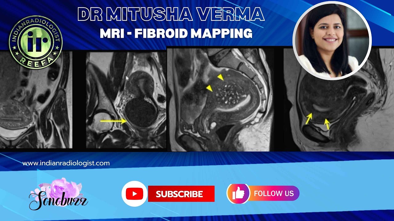
Dr Mitusha Verma | MRI Fibroid Mapping & FIGO classification #mriteachingcourse
Watch on YouTubeVideo summary
Fibroids are now subject to a wide range of treatment options, spanning from non-invasive techniques like MR-guided focused ultrasound (MRgFUS) to invasive surgical procedures such as myomectomy or hysterectomy. In the context of MRI, the large field of view allows clinicians to precisely map the location, number, and proximity of fibroids to adjacent structures. Furthermore, analysis of T2-weighted images and post-contrast dynamic sequences provides critical information regarding vascularity, helping to determine whether a fibroid will respond to MRgFUS or if it requires uterine artery embolization. The most crucial aspect of this imaging is the ability to predict sarcomatous changes within the fibroid, which is essential for guiding management decisions.
The FIGO classification system categorizes fibroids based strictly on their location relative to the uterine wall, a distinction that directly correlates with clinical symptoms. Submucosal fibroids, whether pedunculated or intracavitary, as well as those that are partly submucosal and partly intramural, tend to cause significant issues such as dysmenorrhea, menorrhagia, and infertility due to their pressure on the endometrial cavity. In contrast, purely intramural fibroids generally present fewer symptoms. The classification also includes subserosal types, which may be entirely serosal, partly serosal and intramural, or large enough to indent both the serosa and touch the endometrial cavity, known as type 2-5. Pedunculated subserosal fibroids are also categorized separately, though they can sometimes be confused with broad ligament or wandering fibroids, a confusion that MRI helps resolve by identifying the organ of origin and distinguishing them from ovarian lesions.
On MRI, fibroids typically display a well-defined morphology with a whorled pattern and appear homogeneously iso- to hypointense on T2-weighted sequences. However, as they grow or undergo degeneration, such as hyaline or mixed degeneration, they become heterogeneous; while this heterogeneity is common, it does not necessarily indicate malignancy unless accompanied by other specific signs. The cellular nature of the fibroid influences its signal intensity, with more cellular tumors appearing hypointense on T2, and fat content can be identified using plain T1 sequences followed by fat-suppressed imaging to diagnose lipoleiomyoma. It is vital to differentiate these benign changes from true sarcomatous transformation, which requires careful evaluation beyond just the presence of heterogeneity.
Finally, it is important to distinguish fibroids from adenomyosis, another condition within the endometriosis spectrum that can mimic a fibroid but presents with ill-defined margins rather than the sharp boundaries seen in leiomyomas. Adenomyosis manifests as an asymmetrically bulky uterus where either the anterior or posterior myometrium is thickened. Within this enlarged myometrium, one observes scattered T2 or T1 hyperintense foci representing ectopic endometrial tissue. These areas are relatively well-defined but lack the crisp demarcation of a typical fibroid, making accurate differentiation essential for proper diagnosis and treatment planning in patients presenting with uterine masses.
Read the full video transcript
[music]
>> So, fibroids basically they are now
amenable to a lot of treatment options
from non-invasive like MRgFUS to
practically invasive which is surgery
itself, myomectomy or hysterectomy. So,
what is important MRI wise is the field
of view is very large and we can give
them the exact idea of location of
fibroids, number of fibroids, and their
proximity to various other structures
adjoining it. And the way the fibroid is
appearing on T2,
and post-contrast dynamic sequence, we
can talk about the vascularity of
fibroid, whether it will respond to
MRgFUS or if it will require uterine
artery embolization, or something like
towards the management part part of it.
And most important, if we can predict
any sarcomatous change which has started
within this fibroid, that is the most
important point.
So, basically this FIGO grading is also
available and that is based on the
location of fibroid, nothing else. And
this is what you can put on your reports
also, depending on whether it is a
pedunculated intracavitary fibroid, it's
intramural,
that is one and part of it is
submucosal,
then
part two is again it's more than 50%
intramural and little bit submucosal,
then three which is 100% intramural with
just endometrial contact. So, as we know
the fibroids which are submucosal or
just pressing on the endometrial cavity
are going to cause some symptoms as
compared to those who are completely
intramural. So, dysmenorrhea,
menorrhagia, all those kinds of symptom
are more prone and even infertility with
these kind of submucosal fibroids. So,
that is important.
And then you have the subserosal
fibroid, which is five and
partly subserosal, partly intramural.
When it's a very big fibroid which is
touching the endometrial cavity also and
indenting the serosa also, we call it
two dash five. So, it is both. So,
everything intramural, submucosal,
subserosal.
And then seven is pedunculated
subserosal. These may be confusing
sometime, and MRI definitely helps us to
know the organ of origin because they
may be kind of broad ligament fibroids
or wandering fibroids and also getting
confused with ovarian lesions.
So, this is easy to pick up. These have
like typical appearances, the world
pattern, well-defined morphology. Most
of them are homogeneously T2 iso to hypo
intense, but with increase in size and
starting of degeneration of various
types, like hyaline degeneration and
mixed degeneration, we'll see
heterogeneity within these fibroids. But
only heterogeneity is not anything of
much concern. So, what is important is
to look at other sequences. We'll see
how do we see for the sarcomatous
changes.
So, basically, the degeneration will be
either cystic, hyaline, or mixed. The
most common ones are cystic or hyaline
degeneration, and they start looking
like heterogeneous on T2 weighted
sequences with world pattern and T2
hyperintense areas within.
The more these fibroids are cellular,
they'll be more hypo intense on T2
weighted sequences. They can have fat
content within, so your plain T1 will be
important before you give contrast. And
if that fat component is seen, you have
fat suppressed sequences to prove that
it is fat, and then it becomes a
lipoleiomyoma.
Another part is adenomyosis. We'll see
in endometriosis spectrum also, but
these are similar looking to a fibroid,
but the margins are really ill-defined
as compared to what we saw for fibroids.
And you will have a adenomyotic uterus.
So, asymmetrically bulky uterus where
the anterior or posterior myometrium
will be asymmetrically thick, and these
typical T2 or T1 hyperintense foci will
be seen scattered within the myometrium.
And then a relatively
well-defined area but less well-defined
as compared to a fibroid which will be
present in a adenomyotic uterus.